Pelvic Anatomy / Anatomy Of The Female Pelvis Springerlink : Fold of peritoneum that connects anterior ovary with posterior….. Ct body (lymph nodes) ct. During pregnancy, the pelvic joints and ligaments are relaxed, so that the relaxed, so that the range of motion is increased and the locking mechanism becomes less efficient. • pelvis begins at the iliac crests and ends at the symphysis pubis. Broad ligament of the uterus. The pelvis is the part of the body located between the abdomen and the thighs.
Each hip bone, in turn, is firmly joined to the axial skeleton via its attachment to the sacrum of the vertebral column. The term `pelvis` can refer to the pelvic skeleton (also known as the pelvic girdle), which is the skeleton embedded in the lower part of the trunk, connecting the axial skeleton to the lower extremities. This mechanistic approach should help guide research into pathophysiology, The pelvis is the part of the body located between the abdomen and the thighs. Surgical anatomy of the female pelvis by laparoscopy.
Pelvis And Perineum Anatomy Vessels Nerves Kenhub from thumbor.kenhub.com Johns hopkins medicine, based in baltimore, maryland During pregnancy, the pelvic joints and ligaments are relaxed, so that the relaxed, so that the range of motion is increased and the locking mechanism becomes less efficient. The lining of the uterus. The pelvis is the lower portion of the trunk, located between the abdomen and the lower limbs. It is strengthened and supported by several joints and ligaments. Use the mouse scroll wheel to move the images up and down alternatively use the tiny arrows (>>) on both side of the image to move the images.>>) on both side of the image to move the images. Ilium, ischium, and pubis, meeting in the acetabular fossa at the triradiate fusion center. Pelvic anatomy is composed of two innominate (coxal) bones that articulate with the sacrum and proximal.
The pelvic bones are smaller and narrower.
Anatomy of female pelvic area. Fold of peritoneum that connects anterior ovary with posterior…. Laparoscopic anatomy of the female pelvic region. The pelvis is the lower part of the torso. However, knowledge of the anatomy of various structures that surround these organs has evolved over time. Reviews the functional anatomy of the pelvic floor structures, the effects of age on urethral support and the urethral sphincter, and attempts to clarify what is known about the different structures that influence stress continence. Describe the boundaries and subdivisions of the pelvis. The pelvis is the part of the body located between the abdomen and the thighs. • pelvis begins at the iliac crests and ends at the symphysis pubis. Gross anatomy of the pelvis—namely the bladder, uterus, fallopian tubes, ovaries, rectum, and the muscles—has remained unchanged; The pelvic girdle and pelvic spine. The pelvic girdle (hip girdle) is formed by a single bone, the hip bone or coxal bone (coxal = hip), which serves as the attachment point for each lower limb. The lining of the uterus.
Gross anatomy of the pelvis—namely the bladder, uterus, fallopian tubes, ovaries, rectum, and the muscles—has remained unchanged; Two female reproductive organs located in the pelvis. The pelvis is the lower portion of the trunk, located between the abdomen and the lower limbs. This naturally puts a greater strain on the ligaments. Use the mouse scroll wheel to move the images up and down alternatively use the tiny arrows (>>) on both side of the image to move the images.>>) on both side of the image to move the images.
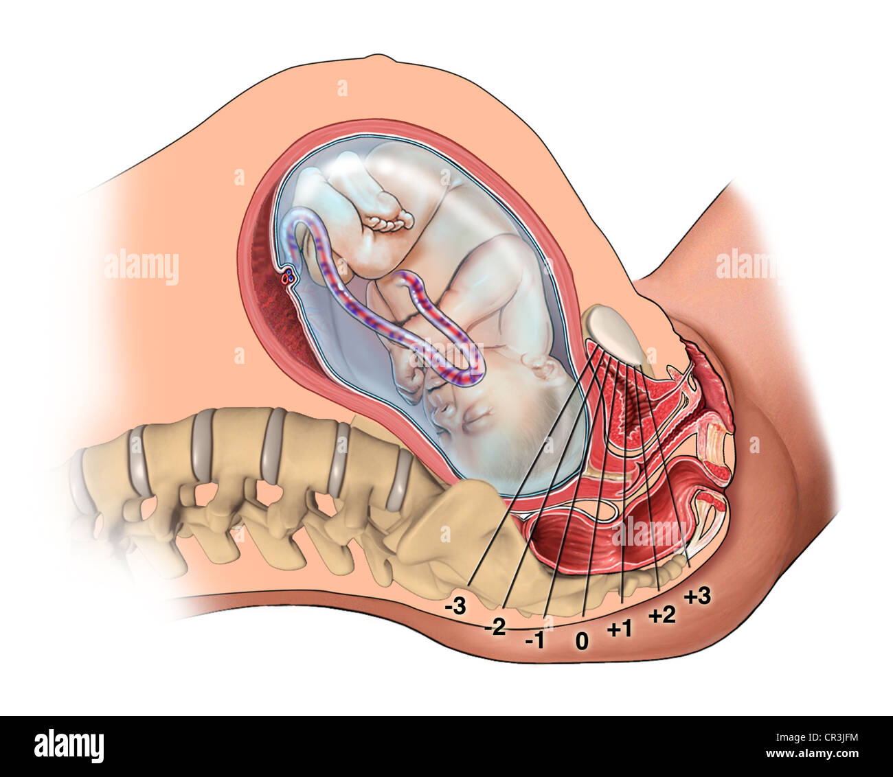
Oa Fetus Presentation Pregnant Female Pelvic Anatomy Stock Photo Alamy from c8.alamy.com It provides attachment to some important muscles in the region, and forms a cavity which accommodates several important internal organs. Space between bladder and uterus. List the arterial & nerve supply list the lymph & venous drainage of the pelvis. Reviews the functional anatomy of the pelvic floor structures, the effects of age on urethral support and the urethral sphincter, and attempts to clarify what is known about the different structures that influence stress continence. The male pelvis is different from a female's. By continuing to browse this site you are agreeing to our use of cookies. Describe the components & function of the pelvic diaphragm. Two female reproductive organs located in the pelvis.
Pelvis (hip) anatomy quiz for anatomy and physiology!
However, knowledge of the anatomy of various structures that surround these organs has evolved over time. Each hip bone, in turn, is firmly joined to the axial skeleton via its attachment to the sacrum of the vertebral column. Differentiate the different types of the female pelvis. This cavity is located within the lesser part of the pelvis, beneath the pelvic brim. Describe the boundaries and subdivisions of the pelvis. The pelvic region is the area between the trunk — or main body — and the lower extremities, or legs. Über 7 millionen englischsprachige bücher. Johns hopkins medicine, based in baltimore, maryland It's located between the abdomen and the legs. Laparoscopic anatomy of the female pelvic region. The pelvic bones are smaller and narrower. Pelvis (hip) anatomy quiz for anatomy and physiology! Two female reproductive organs located in the pelvis.
• divided into the true and false pelvis by the iliopectineal line. Ct body (lymph nodes) ct. Use the mouse scroll wheel to move the images up and down alternatively use the tiny arrows (>>) on both side of the image to move the images.>>) on both side of the image to move the images. The pelvis is the lower part of the torso. Über 7 millionen englischsprachige bücher.
Pelvic Floor Disorders Anatomy Primal Pictures from www.primalpictures.com Über 7 millionen englischsprachige bücher. Describe the components & function of the pelvic diaphragm. This area provides support for the intestines and also contains the bladder and reproductive organs. The pelvis's frame is made up of the bones of the pelvis, which connect the axial skeleton to the femurs, and therefore acts in weight bearing of the upper body. This mechanistic approach should help guide research into pathophysiology, When you are taking anatomy and physiology you will be required to know the anatomical structure locations of the pelvis. The pelvis (plural pelves or pelvises) is either the lower part of the trunk of the human body between the abdomen and the thighs (sometimes also called pelvic region of the trunk) or the skeleton embedded in it (sometimes also called bony pelvis, or pelvic skeleton). Covering a compendium of gynecologic operations, including major and minor procedures and approaches, the techniques.
Covering a compendium of gynecologic operations, including major and minor procedures and approaches, the techniques.
It is further divided into the greater (false) and lesser (true) pelvis. This area provides support for the intestines and also contains the bladder and reproductive organs. The term `pelvis` can refer to the pelvic skeleton (also known as the pelvic girdle), which is the skeleton embedded in the lower part of the trunk, connecting the axial skeleton to the lower extremities. Ct body (lymph nodes) ct. Broad ligament of the uterus. When you are taking anatomy and physiology you will be required to know the anatomical structure locations of the pelvis. The pelvis's frame is made up of the bones of the pelvis, which connect the axial skeleton to the femurs, and therefore acts in weight bearing of the upper body. Complete coverage of both conventional and endoscopic surgeries helps you master the full spectrum of surgical procedures. Johns hopkins medicine, based in baltimore, maryland This quiz is unlabeled so it will test your knowledge on how to identify these structural locations (iliac crest, ischial spine, acetabulum, superior ramus of pubis, posterior superior/inferior iliac spine, lessier. Anatomy of female pelvic area. Über 7 millionen englischsprachige bücher. Use the mouse scroll wheel to move the images up and down alternatively use the tiny arrows (>>) on both side of the image to move the images.>>) on both side of the image to move the images.


0 Komentar